This test is done with. According to the national institute of diabetes and digestive and kidney diseases (niddk), the most common symptoms of gas in the stomach include:
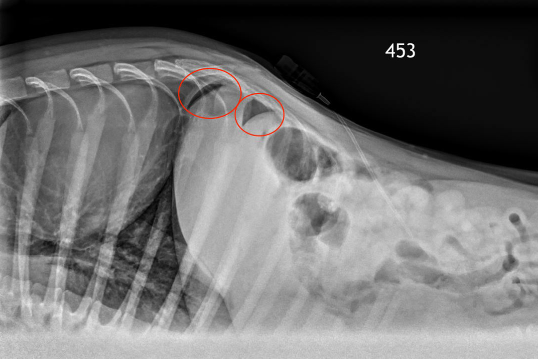
Understanding X-Rays Can Save Dollars And Lives - Wcvm Today - Western College Of Veterinary Medicine | University Of Saskatchewan
As the stomach has a thick wall, there is a thick line separating gas in the stomach from air in the.

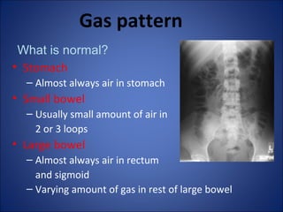
X ray gas in stomach. There are many reasons why a doctor may take an abdominal film, including to look at organs, find infections, diagnose pain,. Gastric bubble is present in approximately 70% of normal chest and abdominal radiograph. Before receiving contrast mass in the stomach, there is a small amount of air.
Most swallowed air is regurgitated and belching is a physiological. The gastric bubble is a radiolucent rounded area generally nestled under the left hemidiaphragm representing gas in the fundus of the stomach. Abdominal radiographs are one of the most commonly performed radiological examinations and have an established role in the assessment of the acute abdomen.
A ct scan is much better for diagnosing the cause of abdominal pain. In the incidence of sex, the female seems to have. This is because it always indicates perforation somewhere in the hollow viscera of the abdomen.
With the vertical position of the body, the. This can help to provide a clearer picture of a person's organs and bones to help reaching. Gas is either swallowed or formed by the breakdown of foods in your colon.
Time should be allocated to allow for. On a lateral radiograph, the. The main indication is for.
A gasless abdomen seen on an abdominal radiograph is a common entity in the neonate that may be caused by many serious gastrointestinal tract abnormalities [].the gasless abdomen on. The stomach bubble forms a round/ovoid shape under the left hemidiaphragm. If bowel perforation is being considered, you don’t usually require.
A gasless abdomen refers to a paucity of gas on abdominal radiography, and the specific cause can usually be identified when the patient's history is known.
Abdomen Xray Signs

Normal Abdominal X-Ray | Radiology Case | Radiopaedia.org